Welcome to Krishnam Womens Hospital

  • Emergancy Helpline: +91-840 181 2598, 840 191 2598

Ultra Sonography

Awesome Image


What is ULTRA SONOGRAPHY??

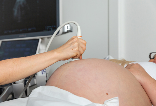
Ultrasound, or ultrasonography, is a medical imaging technique that uses high-frequency sound waves to create images of the inside of the body. It's like taking a picture, but instead of using light, it uses sound. A small device called a transducer sends out these sound waves into the body, which then bounce back when they hit organs, tissues, or other structures. The returning sound waves are captured by the transducer and converted into images that can be viewed on a screen. These images help doctors to see the internal organs and structures in real-time, allowing them to diagnose and monitor various medical conditions without the need for invasive procedures. Ultrasound is commonly used during pregnancy to monitor the development of the fetus, but it can also be used to examine organs such as the heart, liver, kidneys, and reproductive organs. It's safe, painless, and doesn't involve radiation, making it a preferred choice for many medical situations.

Awesome Image


What is NT NB SCAN??

Nuchal Translucency and Nasal Bone Scan, , , is a prenatal screening procedure used to assess the risk of chromosomal abnormalities in a developing fetus, particularly Down syndrome (Trisomy 21), Edwards syndrome (Trisomy 18), and Patau syndrome (Trisomy 13). During this non-invasive ultrasound scan, the thickness of the nuchal translucency (the fluid-filled space at the back of the fetus's neck) and the presence or absence of the nasal bone are measured. An increased thickness of the nuchal translucency or absence of the nasal bone may indicate a higher risk of chromosomal abnormalities. NT NB SCAN is typically performed between 11 and 14 weeks of pregnancy and is often combined with blood tests for a more comprehensive assessment of the fetus's health. It helps expectant parents and healthcare providers make informed decisions regarding further diagnostic testing or intervention if necessary.

Awesome Image


What is A TIFFA SCAN??

TIFFA scan is , Targeted Imaging for Fetal Anomalies, is like a detailed check-up for a baby still growing inside its mom's belly. It's usually done around 18 to 22 weeks into the pregnancy. During the scan, a special machine uses sound waves to take pictures of the baby. The doctor looks closely at these pictures to make sure everything is developing as it should be. They check the baby's brain, spine, heart, and other parts to see if there are any problems or things that need extra attention. If they find anything concerning, they may suggest more tests or treatments to keep both the baby and the mom healthy. Overall, the TIFFA scan helps doctors keep an eye on how the baby is doing before it's born..

Awesome Image


What is a 3D - 4D SONOGRAPY??

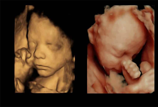
3D-4D sonography , is like a special photo shoot for babies before they're born. Instead of traditional ultrasound pictures, which are flat, this scan creates 3D images, so you can see the baby's features more clearly, almost like a photograph. The "4D" part means it's like watching a live video because it shows the baby moving in real-time. It's usually done during pregnancy to check on the baby's growth and development. Doctors use it to see if everything is okay with the baby's organs, limbs, and other body parts. Parents often love these scans because they get a better look at their little one before they arrive. Overall, it's a cool way to bond with the baby and make sure everything's going smoothly during pregnancy.

Awesome Image


What is a COLOUR DOPPLER SCAN ?

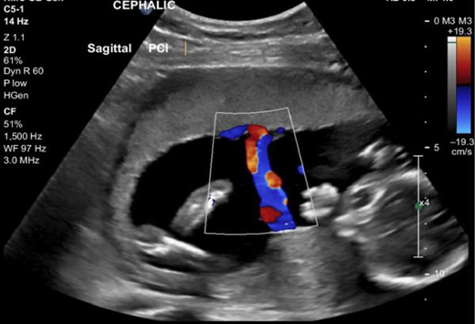
A Colour Doppler , scan is a special type of ultrasound that helps doctors see how blood is flowing through the body's blood vessels. It's like looking at a map where different colors represent the direction and speed of blood flow. This scan is often used to check the blood flow in the arteries and veins, helping doctors diagnose conditions like blood clots, blockages, or problems with the heart or other organs. During the scan, a gel is applied to the skin, and a device called a transducer is gently moved over the area being examined. It's painless and safe, and it gives doctors important information to help them understand what's happening inside the body. Overall, the Color Doppler scan is a helpful tool for doctors to see how blood is moving around and to spot any issues that need attention.

Awesome Image


What is a GROWTH SCAN??

A growth scan , is like a special check-up for a baby still growing inside its mom's belly. It's done during pregnancy to see how the baby is growing and developing. The scan uses ultrasound to measure the baby's size, weight, and other important details. Doctors use this information to make sure the baby is growing at a healthy rate. They also check the amount of amniotic fluid around the baby, which is like a cushion to keep it safe. If the scan shows any concerns, the doctor may suggest more tests or treatments to help the baby grow better. Overall, the growth scan helps parents and doctors keep track of how the baby is doing before it's born.

Awesome Image


What is a PELVIC SONOGRAPHY??

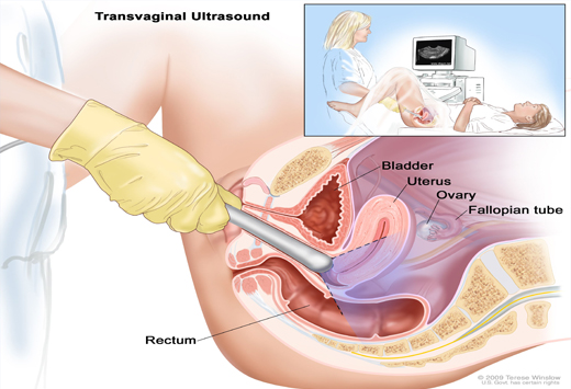
Pelvic sonography ,is a type of ultrasound scan that looks at the organs inside a person's pelvis. It's like taking a picture of the uterus, ovaries, and nearby organs using sound waves. This scan is often used to check for problems like cysts, fibroids, or tumors in the pelvic area. It's also helpful for monitoring pregnancies to make sure the baby is growing properly in the uterus. During the scan, a gel is applied to the skin, and a device called a transducer is gently moved over the lower belly. It's painless and safe, and it gives doctors important information to help them diagnose and treat pelvic conditions. Overall, pelvic sonography is a valuable tool for doctors to see inside the pelvis and find any issues that need attention.